Dec
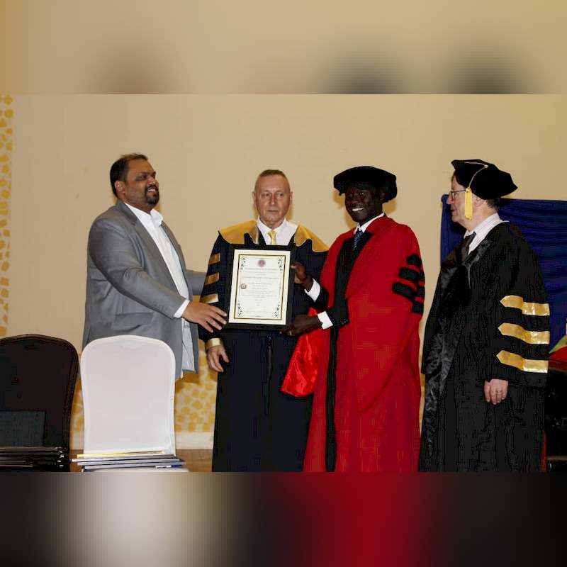
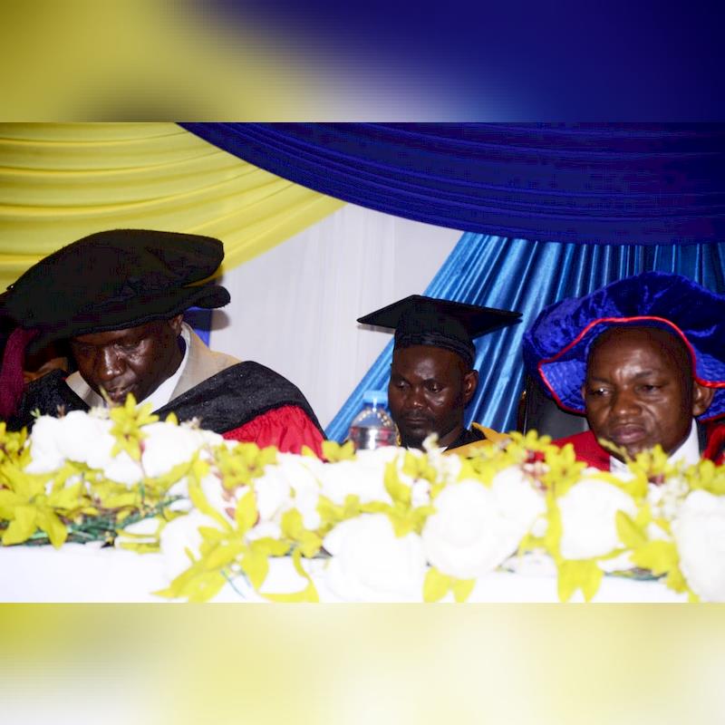
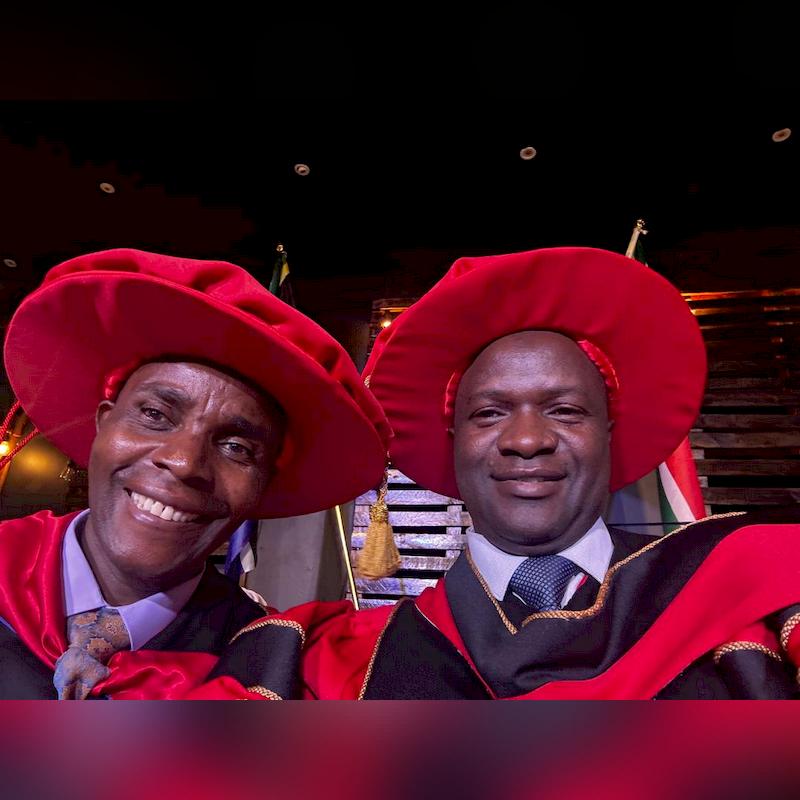
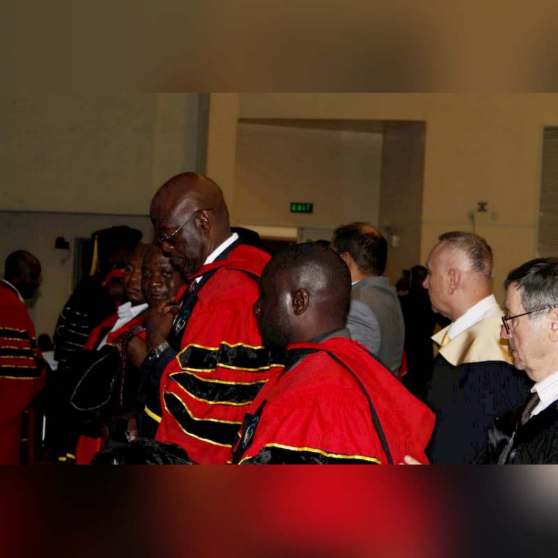
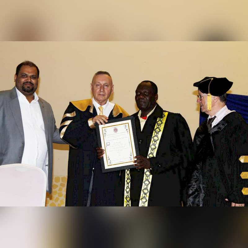
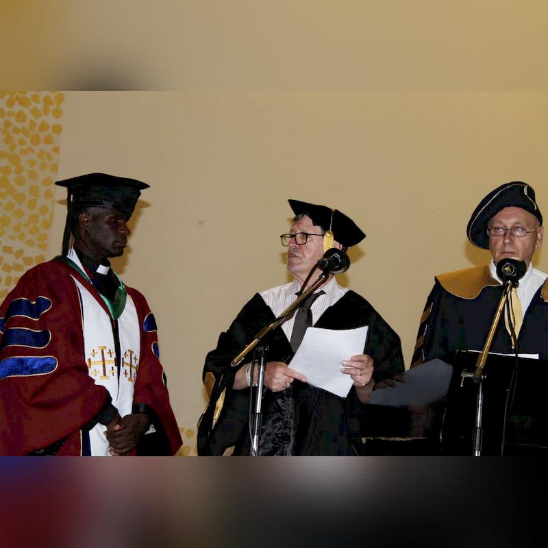
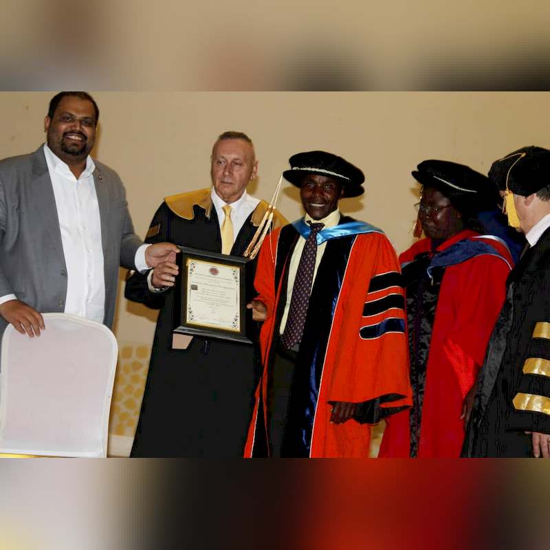
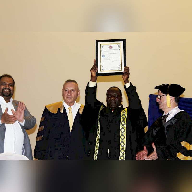
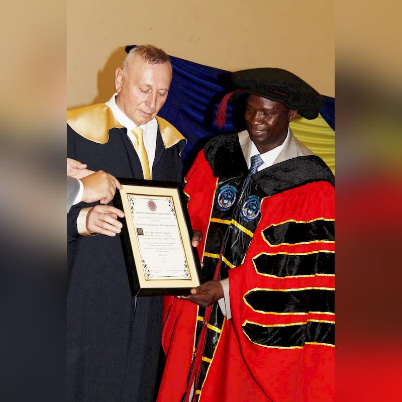
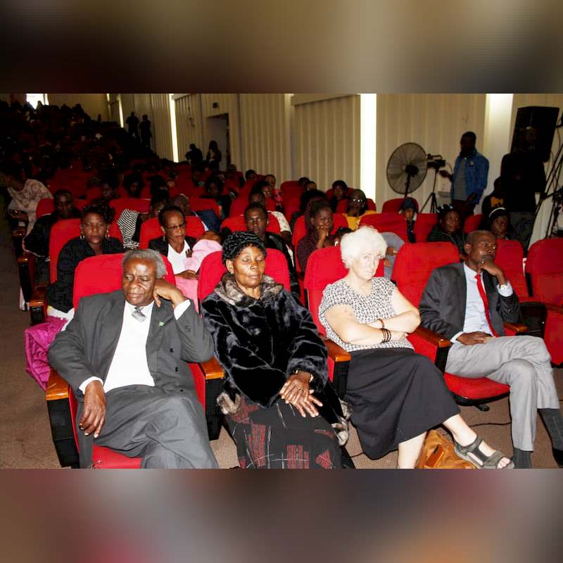
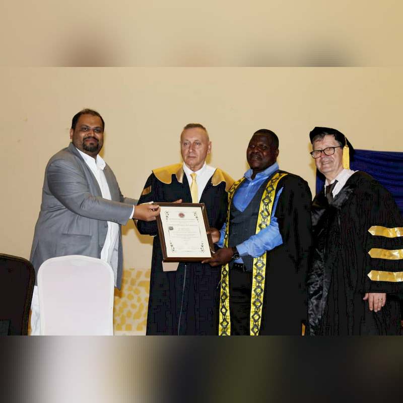
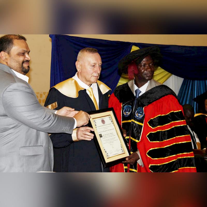
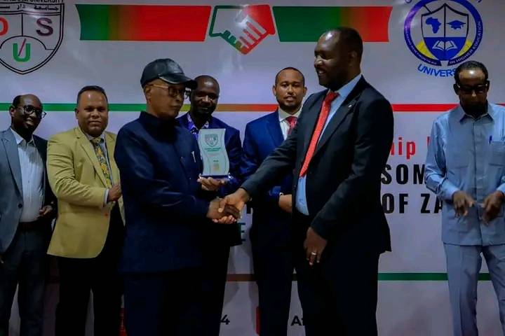
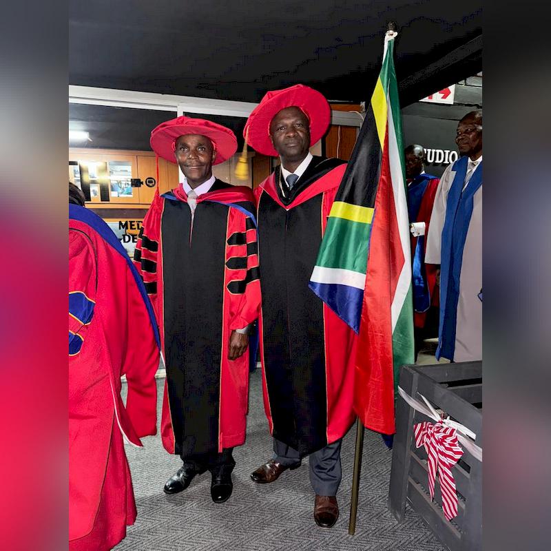
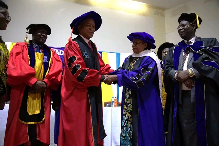
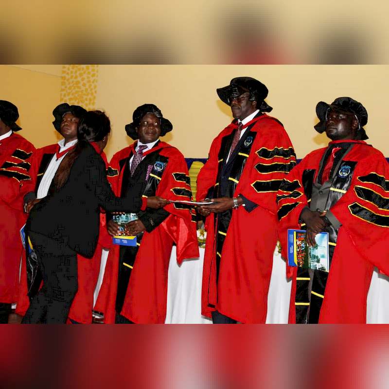
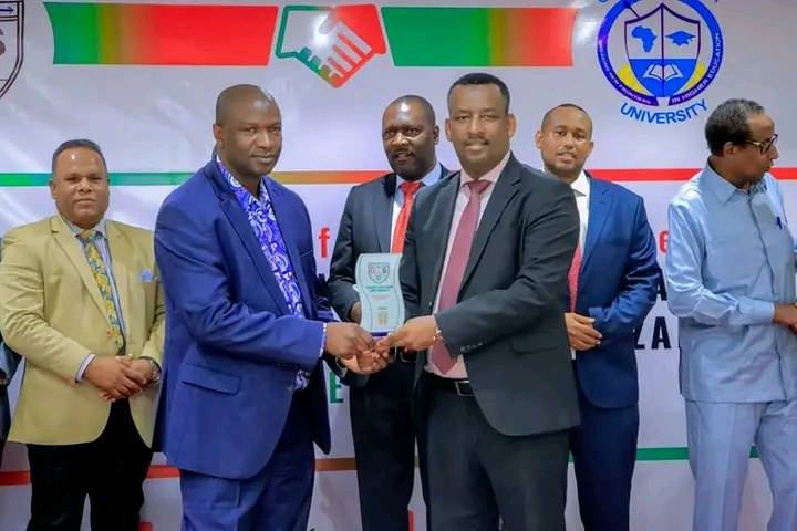
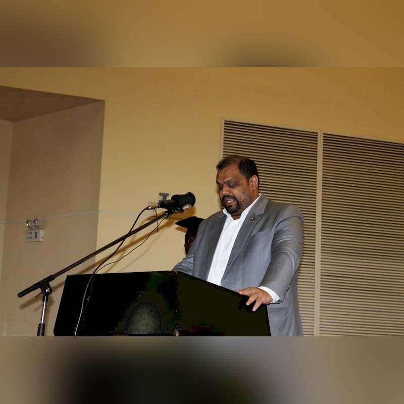
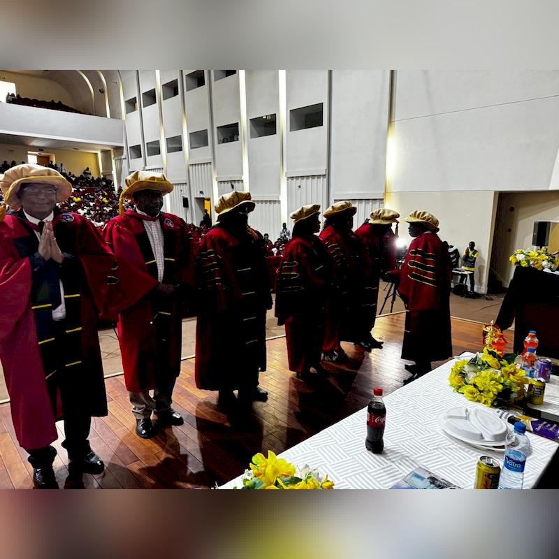
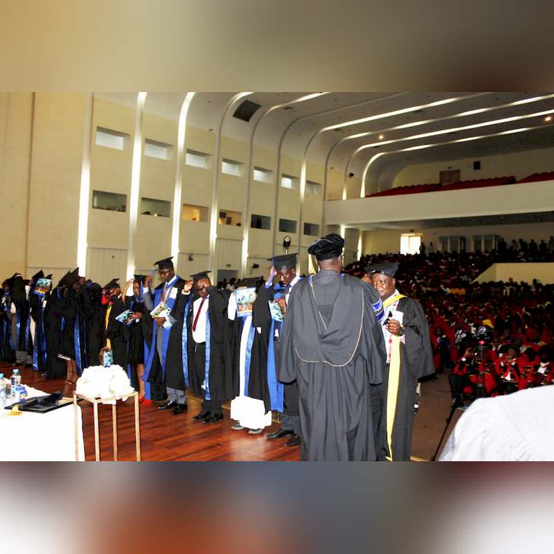
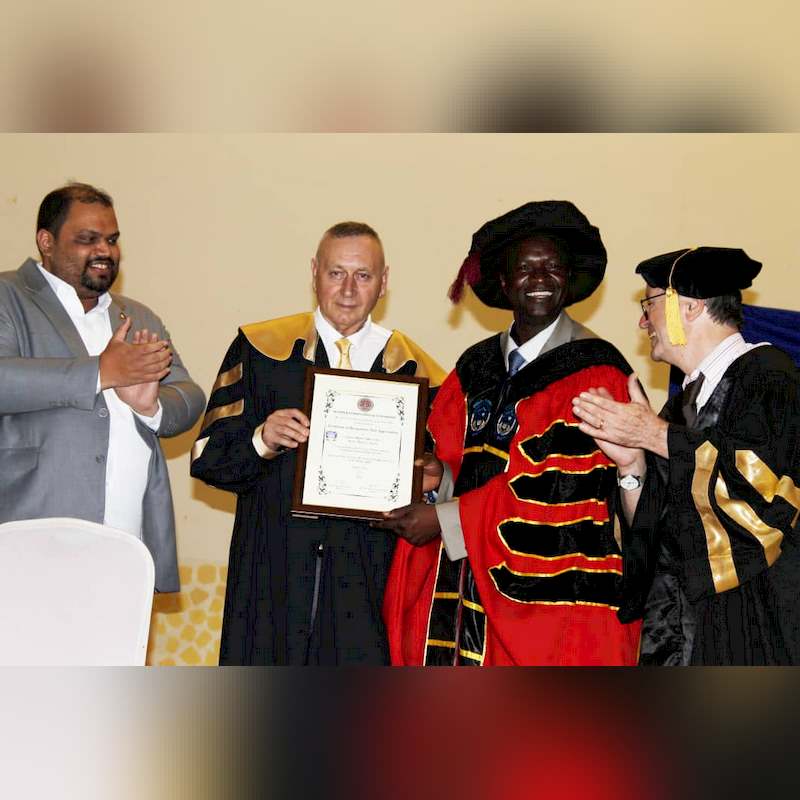
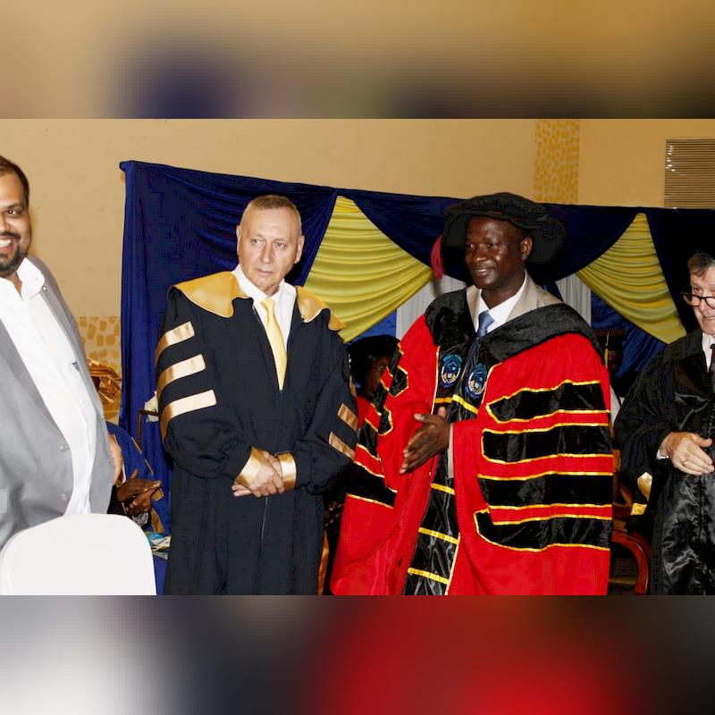
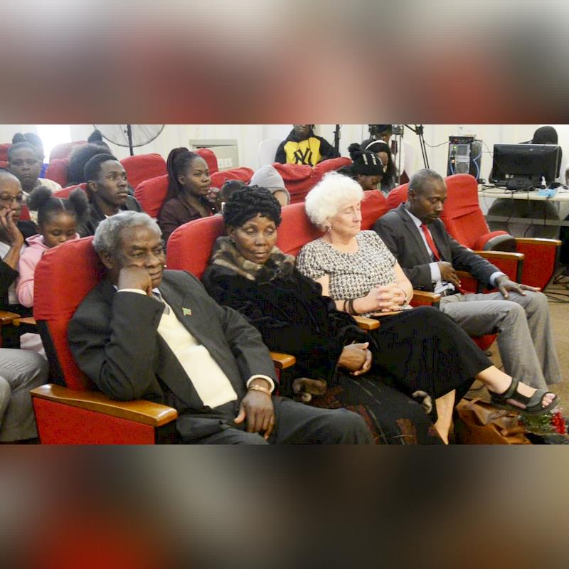
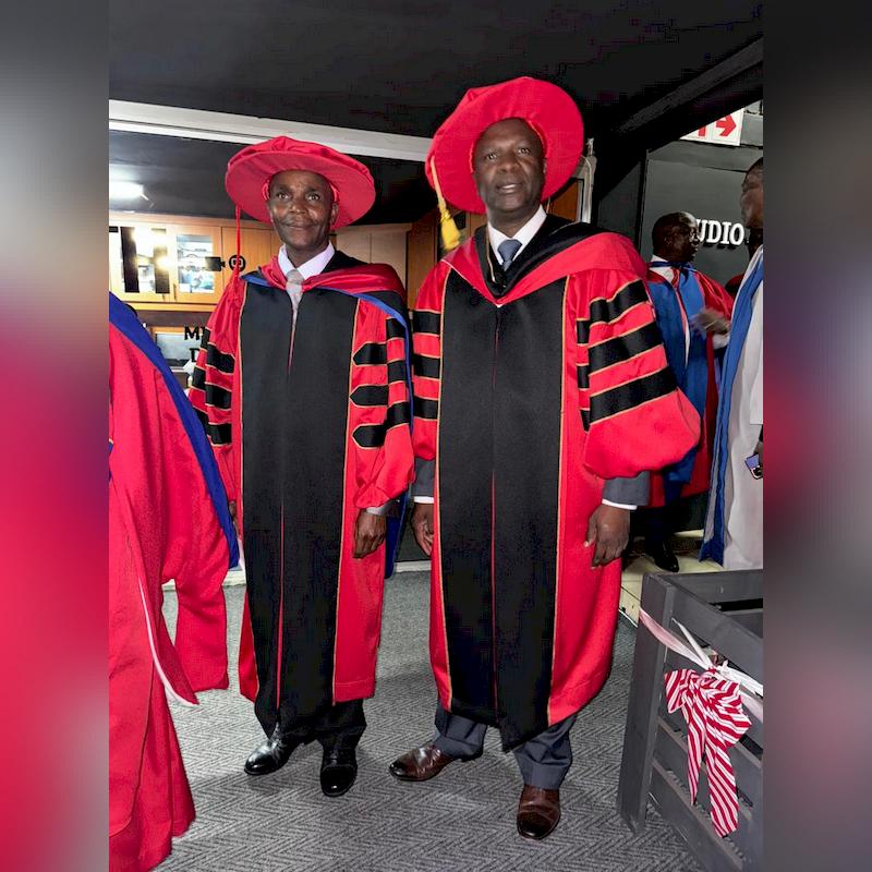
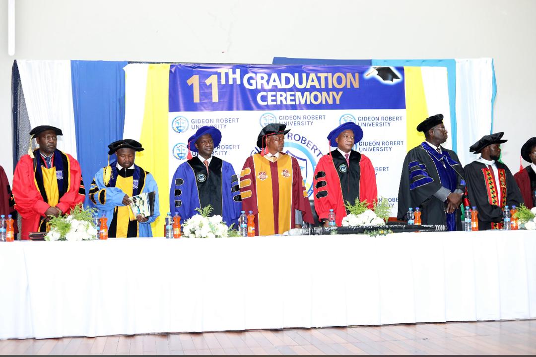
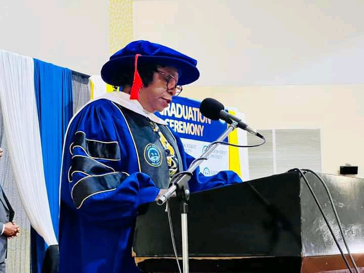
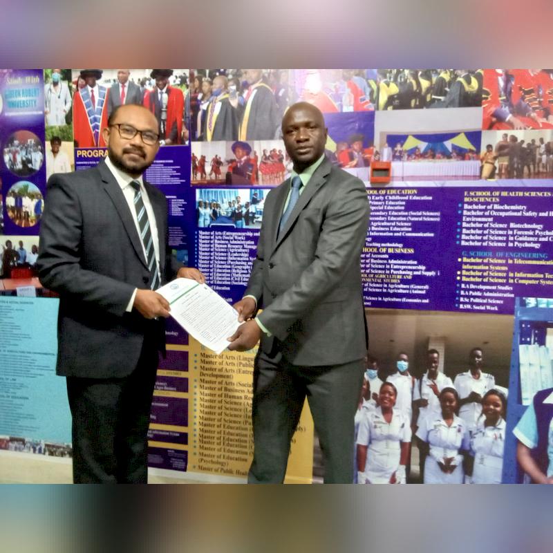
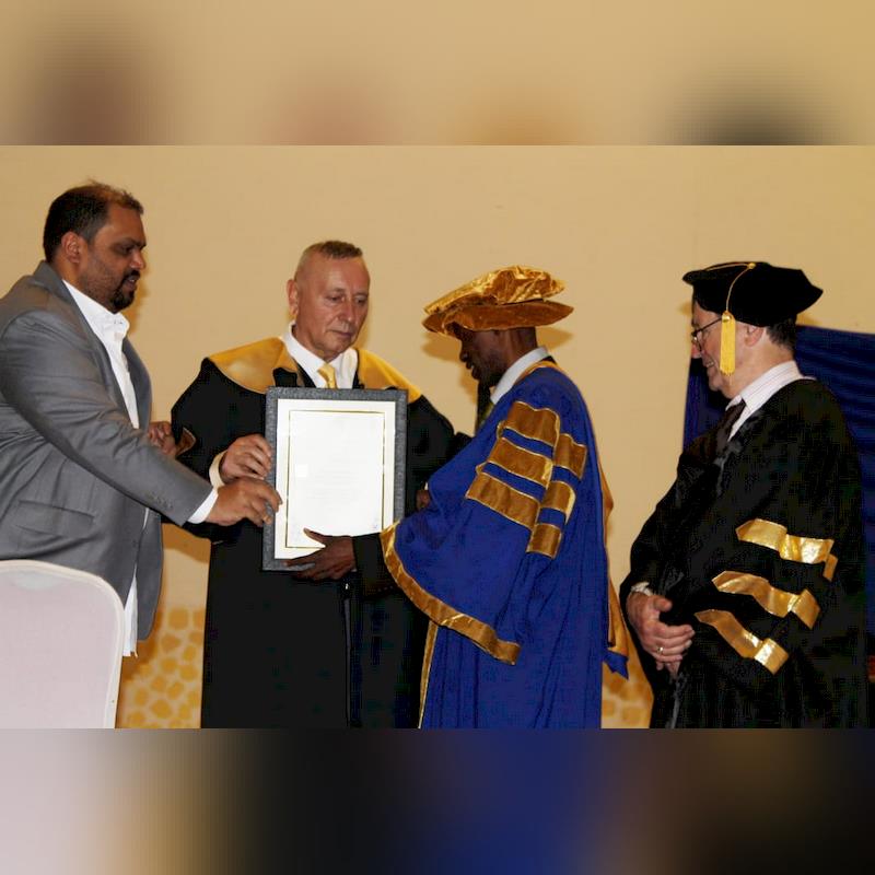
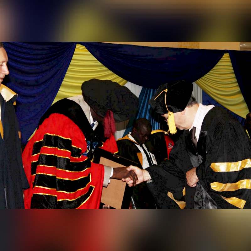
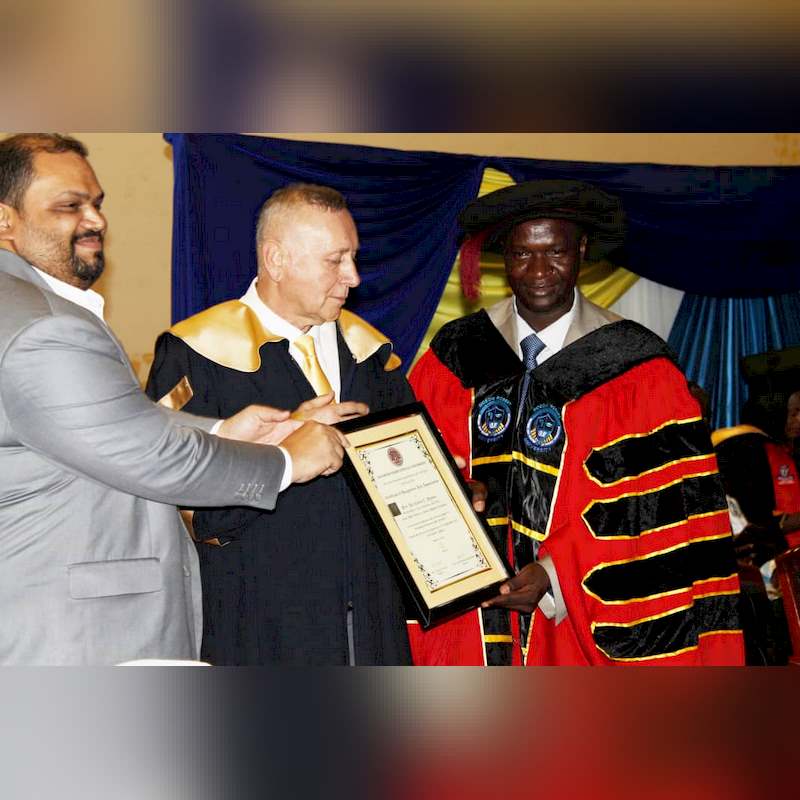
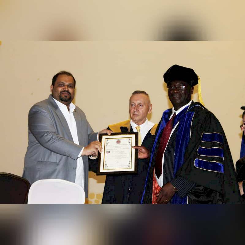
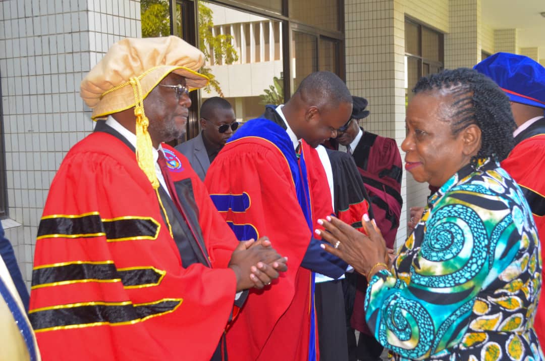
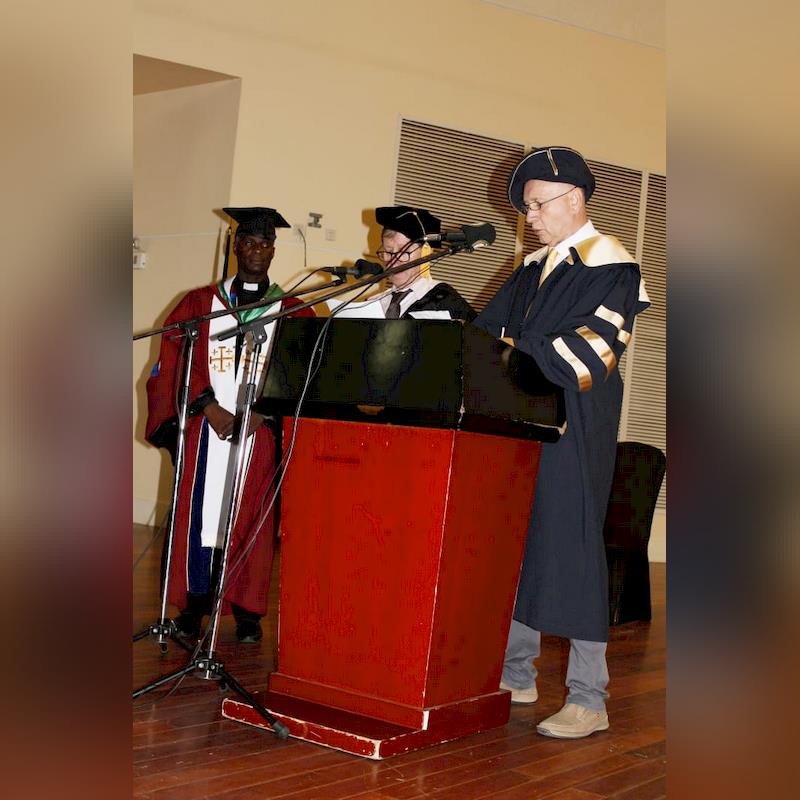
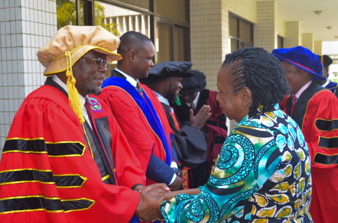
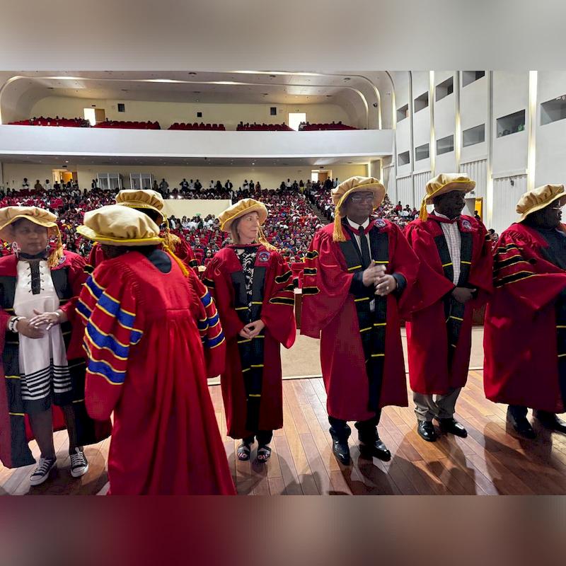
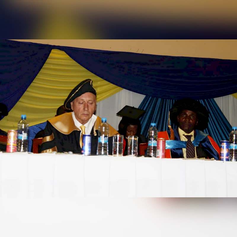
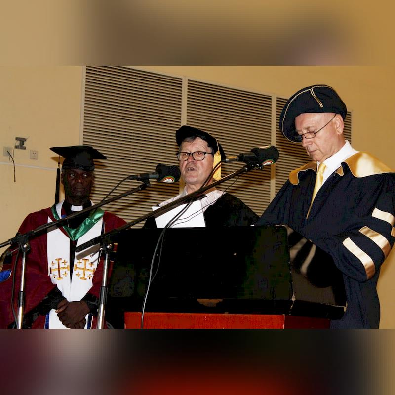
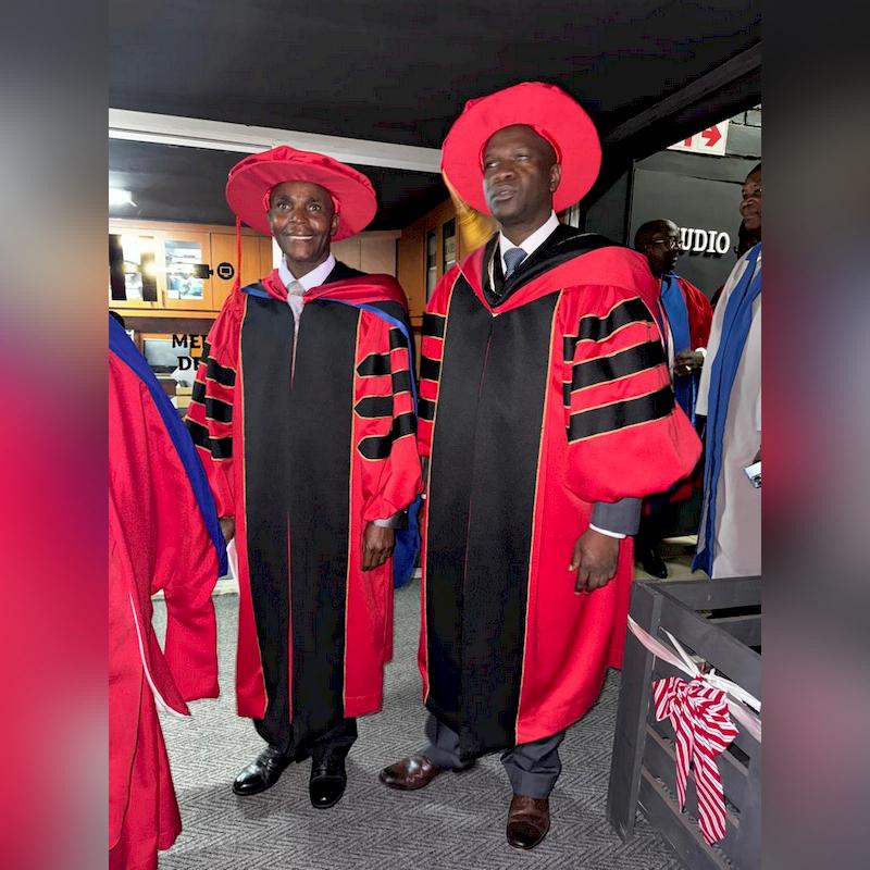
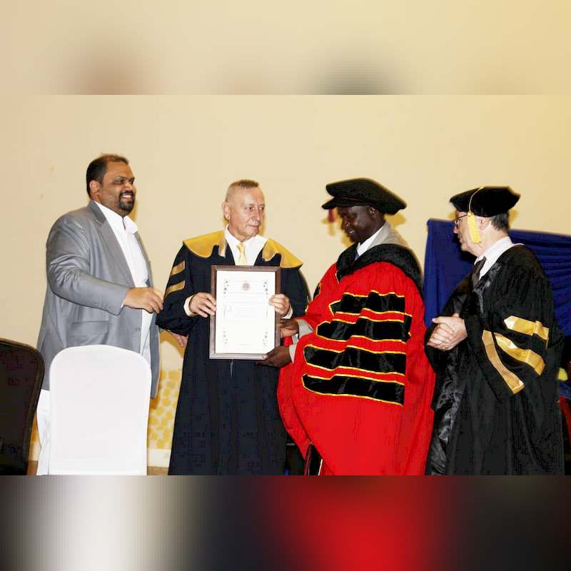
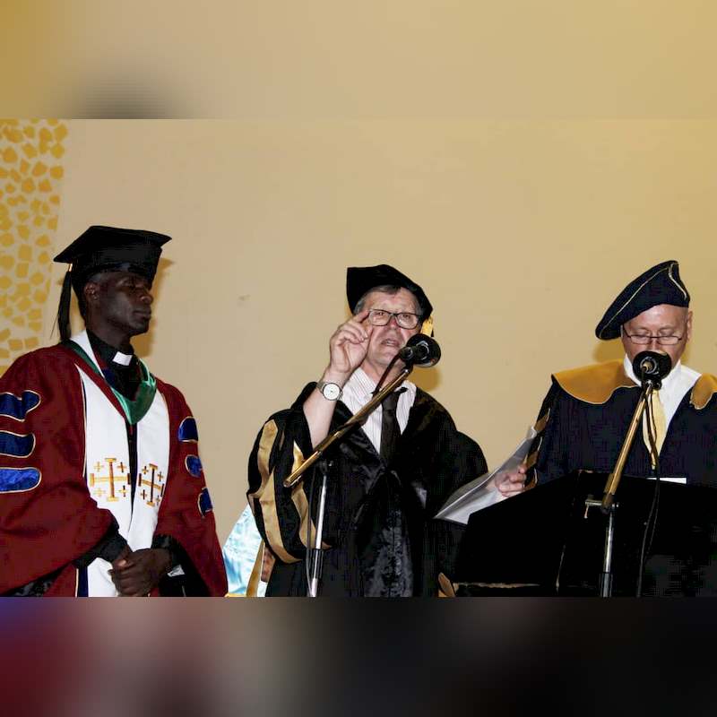
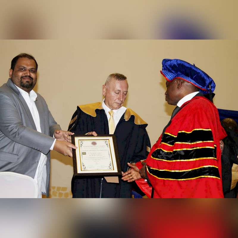
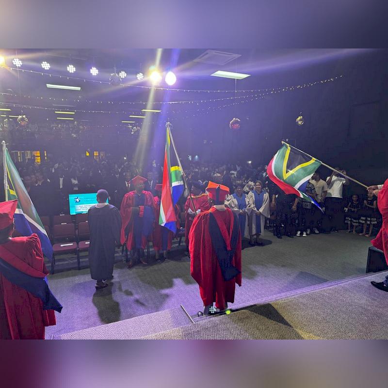
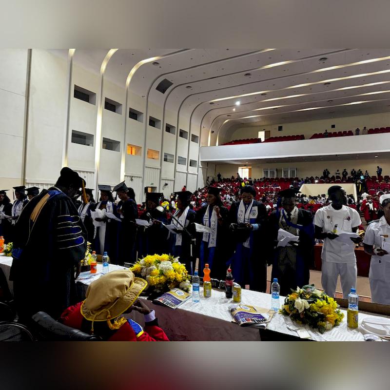
2024VICE PRESIDENT W.K MUTALE-NALUMANGO APPLAUDS THE POSITIVE IMPACT OF EDUCATION ON Society.... Conferred with Honorary doctorate. Vice President Dr. W.K Mutale-Nalumango has commended President Hakainde Hichilema for his unweaving leadership and dedication to the education sector. The Vice President said the deliberate channeling of resources to the education sector has seen increased number of children return to school a focus that is transforming the country. Dr. Nalumango was speaking in Lusaka in Lusaka on Friday 20th December 2024 after she was awarded with Two (2) Honorary doctorate degrees in Education and Political Science from Gideon Roberts University of Zambia and Kesmonds International University of Cameroon respectively. The two degrees were awarded to the Vice President at the 11th Graduation Ceremony of the Gideon Roberts University. The Graduation ceremony was held under the theme: "Empowering future leaders: celebrating academic excellence in career development." The Vic......
Dec
2024......
Nov
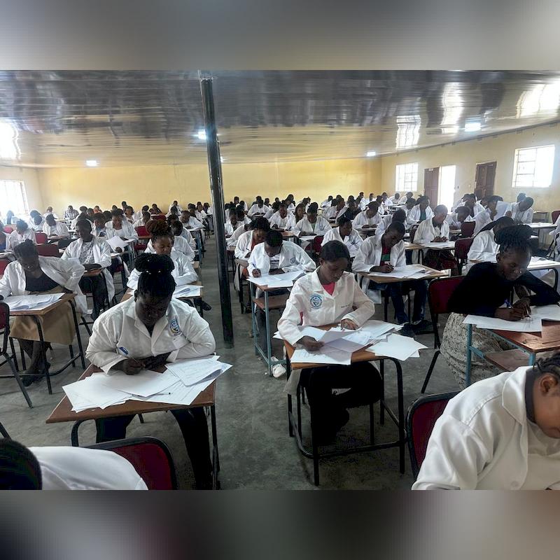
2024EXAMINATION TIME TABLE FOR FULL-TIME AND PART-TIME STUDENTS (2024 NOVEMBER EXAMINATIONS)......