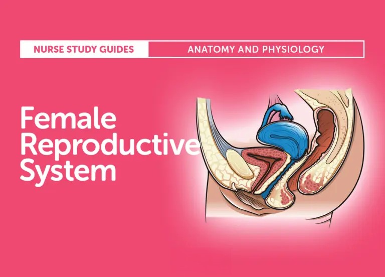
FEMALE REPRODUCTIVE SYSTEM ANATOMY AND PHYSIOLOGY
Female Reproductive System Anatomy and Physiology
Women have the responsibility of bringing forth life into the world, hence the creation and the function of the female reproductive system. This system performs a miracle from the conception of life until the birth of the growing life within, and it is only proper to be introduced to the main characters and supporting roles of this play.
Internal Structures
Ovaries
- The ovaries are the ultimate life-maker for the females.
- For its physical structure, it has an estimated length of 4 cm and width of 2 cm and is 1.5 cm thick. It appears to be shaped like an almond. It looks pitted, like a raisin, but is grayish white in color.
- It is located proximal to both sides of the uterus at the lower abdomen.
- For its function, the ovaries produce, mature, and discharge the egg cells or ova.
- Ovarian function is for the maturation and maintenance of the secondary sex characteristics in females.
- It also has three divisions: the protective layer of epithelium, the cortex, and the central medulla.
Fallopian Tubes
- The fallopian tubes serve as the pathway of the egg cells towards the uterus.
- It is a smooth, hollow tunnel that is divided into four parts: the interstitial, which is 1 cm in length; the isthmus, which is2 cm in length; the ampulla, which is 5 cm in length; and the infundibular, which is 2 cm long and shaped like a funnel.
- The funnel has small hairs called the fimbria that propel the ovum into the fallopian tube.
- The fallopian tube is lined with mucous membrane, and underneath is the connective tissue and the muscle layer.
- The muscle layer is responsible for the peristaltic movements that propel the ovum forward.
- The distal ends of the fallopian tubes are open, making a pathway for conception to occur.
Uterus
- The uterus is described as a hollow, muscular, pear-shaped organ.
- It is located at the lower pelvis, which is posterior to the bladder and anterior to the rectum.
- The uterus has an estimated length of 5 to 7 cm and width of 5 cm. it is 2.5 cm deep in its widest part.
- For non-pregnant women, it is approximately 60g in weight.
- Its function is to receive the ovum from the fallopian tube and provide a place for implantation and nourishment.
- It also gives protection for the growing fetus.
- It is divided into three: the body, the isthmus, and the cervix. f
- The body forms the bulk of the uterus, being the uppermost part. This is also the part that expands to accommodate the growing fetus.
- The isthmus is just a short connection between the body and the cervix. This is the portion that is cut during a cesarean section.
- The cervix lies halfway above the vagina, and the other half extends into the vagina. It has an internal and external cervical os, which is the opening into the cervical canal.
External Structures
Mons Veneris
- The mons veneris is a pad of fat tissues over the symphysis pubis.
- It has a covering of coarse, curly hairs, the pubic hair.
- It protects the pubic bone from trauma.
Labia Minora
- The labia minora is a spread of two connective tissue folds that are pinkish in color.
- The internal surface is composed of mucous membrane and the external surface is skin.
- It contains sebaceous glands all over the area.
Labia Majora
- Lateral to the labia minora are two folds of fat tissue covered by loose connective tissue and epithelium, the labia majora.
- Its function is to protect the external genitalia and the distal urethra and vagina from trauma.
- It is covered in pubic hair that serves as additional protection against harmful bacteria that may enter the structure.
Vestibule
- It is a smooth, flattened surface inside the labia wherein the openings to the urethra and the vagina arise.
Clitoris
- The clitoris is a small, circular organ of erectile tissue at the front of the labia minora.
- The prepuce, a fold of skin, serves as its covering.
- This is the center for sexual arousal and pleasure for females because it is highly sensitive to touch and temperature.
Skene’s Glands
- Also called as paraurethral glands, they are found lateral to the urethral meatus and have ducts that open into the urethra.
- The secretions from this gland lubricate the external genitalia during coitus.
Bartholin’s Gland
- Also called bulbovaginal gland, this is another gland responsible for the lubrication of the external genitalia during coitus.
- It has ducts that open into the distal vagina.
- Both of these glands secretions are alkaline to help the sperm survive in the vagina.
Fourchette
- This is a ridge of tissue which is formed by the posterior joining of the labia minora and majora.
- During episiotomy, this is the tissue that is cut to enlarge the vaginal opening.
Perineal Body
- This is a muscular area that stretches easily during childbirth.
- Most pregnancy exercises such as Kegel’s and squatting are done to strengthen the perineal body to allow easier expansion during childbirth and avoid tearing the tissue.
Hymen
- This covers the opening of the vagina.
- It is tough, elastic, semicircle tissue torn during the first sexual intercourse.
Comments